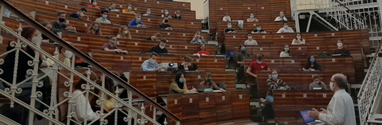
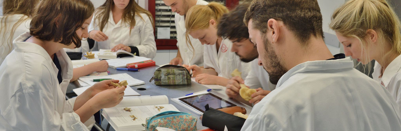
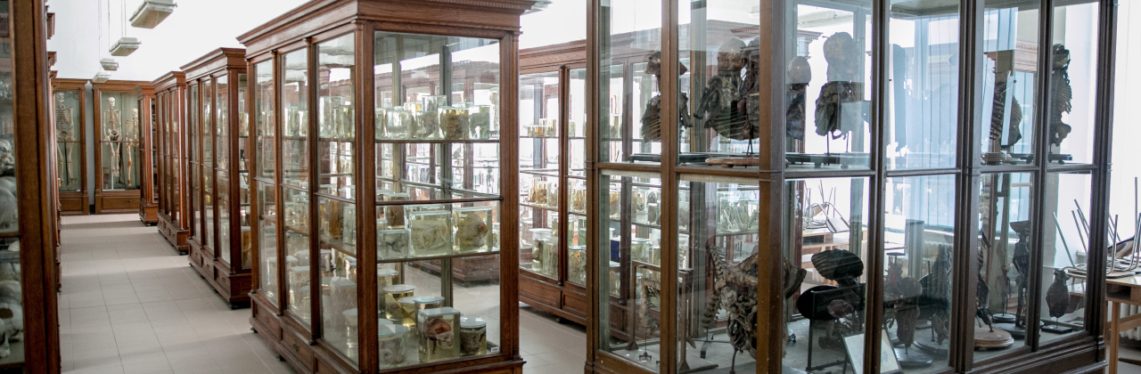
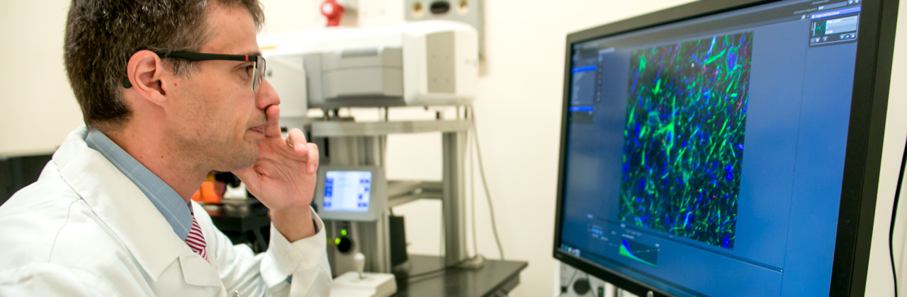
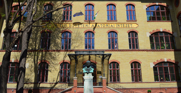
ÖSSZEHASONLÍTÓ NEUROHISZTOLÓGIAI ÉS NEUROCITOKÉMIAI LABORATÓRIUM

Vezető: Dr. Csillag András és Dr. Kálmán Mihály

Név

Beosztás

Tel. E-mail
Dr. Csillag András egyetemi tanár 3681 csillag@ana.sote.hu
Dr. Kálmán Mihály egyetemi docens 3695 kalman@ana.sote.hu
Dr. Székely Andrea Dorottya egyetemi adjunktus 3601 adszekely@ana.sote.hu
dr. Catherine M. Montagnese tudományos munkatárs 3644  
Szász Istvánné asszisztens 3687  
Ari Csilla PhD hallgató (KM) 3626 csari2000@yahoo.com
Mezey Szilvia PhD hallgató (CsA) 3687 mezey@ana.sote.hu
dr. Papp Andrea PhD hallgató(CsA) 3687 pandrea@dec001.geobio.elte.hu
dr. Szabó Adrienn PhD hallgató (KM) 3687 szaboa@ana.sote.hu
Adorján István tudományos diákkörös (KM) 3687 adorist@freemail.hu
Ádám Ágota tudományos diákkörös (CsA) 3687 cicunyatka@yahoo.com
Bálint Eszter tudományos diákkörös (CsA) 3687 pupalibera@hotmail.com
Goren, Oded tudományos diákkörös (KM) 3687 oded_goren@hotmail.com
Kaiser Csilla tudományos diákkörös (KM) 3687  
Kitka Tamás tudományos diákkörös (CsA) 3687 pupalibera@hotmail.com
Kovács Adrienn tudományos diákkörös (CsA) 3687 mississippi@freemail.hu


Laboratóriumunk főként a madarak tanulásában érintett agyterületek és agypályák vizsgálatával, a tanulás során bekövetkező agyi változások követésével foglalkozik.


Kollaborációk

Publikációk


Azonosított striato-ventrotegmentalis projekciós neuronok fény- és elektronmikroszkópos jellemzése a házi csirkében
(Gallus domesticus)

Mezey Szilvia és Csillag András

A medialis striatum egyik legjelentősebb efferentációja a substantia nigrába és az area ventralis tegmentalisba (AVT) vetítő striatotegmentalis pálya. A jelen vizsgálatban az LPO-ban a ventriculus lateralis mentén elhelyezkedő striato-ventrotegmentalis neuronok morfológiai jellemzését végeztük. Az idegsejtek azonosítását az AVT-be injektált fluoreszcens retrográd pályakövető anyag, fast blue (FB) segítségével végeztük. A FB-jelölt neuronokat paraformaldehiddel fixált szeletekben fluoreszcens fénymikroszkóp alatt vizualizáltuk, majd egyes sejteket iontoforézissal célzottan feltöltöttünk fluoreszcens lucifer yellow (LY) festékkel. A töltött sejtekben a LY jelölést fotokonvertáltuk, majd az adott neuronok ultrastruktúráját elektronmikroszkóppal vizsgáltuk.

Eredményeink alapján a striato-ventrotegmentalis neuronok a sejtttestből kiinduló, viszonylag kevés dendrittel rendelkeznek. A sejttest organellumokban gazdag - különösen sima és durva endoplazmatikus retikulumban -, és a nagy sejtmag enyhén excentrikus elhelyezkedésű. Megfigyelhetőek voltak glia közbeiktatása nélküli, közvetlen kapcsolatok a FB-jelölt neuronok és szomszédos neuronok sejttestjei között. a A FB-jelölt neuronokon helyenként axosomaticus szinapszisok voltak láthatóak. A LY jelölés a dendritikus elágazásokban egészen a terminális szegmentumokig követhető volt. Az LY-jelölt dendriteken rövid nyakkal és tömzsi fejjel rendelkező tüskék voltak találhatók. A tüskék nem sűrű elhelyezkedésűek és helyenként hosszú nyakkal és kis fejjel rendelkeznek. A tüskéknek mind a nyakukon, mind a fejükön végtalálhatók szinapszisok, melyek közül a fejen inkább aszimmetrikus szinapszisok találhatók, melyek morfológiailag az LPO-ban korábban leírt glutamáterg, serkentő szinapszisoknak felelnek meg. Az itt leírt idegsejt-típus a mind az emlősök, mind a madarak striatumára jellemző közepes méretű, tüskés neuronoknak felel meg.


Postembrionális neurogenesis vizsgálata, az újjonnan képződött neurononok morfológiai elemzése házi csirkében (Gallus domesticus)

Mind a fiatal és felnőtt állatokban kimutatták új neuronok létrejöttét az agy bizonyos területein. Laboratóriumunkban fiatal házicsirkék subventricularis és paraventricularis zónájában kialakuló neuronok morfológiájának meghatározásával foglalkozunk. Az osztódó neuronokat bromodeoxi-uridinnel megjelöljük, majd az így azonosított neuronok morfológiáját egysejt-feltöltés segítségével, fény- és elektronmikroszóppal vizsgáljuk.


BrdU-jelölt neuronok 3 napos csirke előagyában, 2 nappal a BrdU beadását követően. Az újonnan képződött sejtek a ventriculus lateralis (VL) mentén képződnek és 1 nappal a beadást követően még jól láthatóan ott is tömörülnek.


Résztvevők:

Dr. Kálmán Mihály, egyetemi docens, az orvostudomány doktora

Dr. Szabó Adrienn orvos, másodéves PhD-hallgató

Ari Csilla zoológus, elsőéves PhD-hallgató

Adorján István negyedéves orvostanhallgató

Kajzer Csilla negyedéves orvostanhallgató

Oren Goded harmadéves orvostanhallgató.

Kutatási területek:

    1) Agyi sérülések utáni gliareakciók és esetleges regenerációs jelenségek vizsgálata, kifejlett és fejlődő agyszöveten, nem-emlős gerinceseken is (madáron, hüllőn, csontos- és porcoshalon). Emlősök esetében a vizsgálatokat intrauterin végzett léziókkal a magzati korra is kiterjesztettük. Sikerült találnunk olyan modelleket, ahol a sérülést követő reaktív gliózis az általánosan elfogadott nézettel szemben nem gátolja, sőt, valószínűleg serkenti az idegrostok növekedését.

    2) A glia és a kötőszövet kapcsolata, ennek jelentősége az agy fejlődésében, vaszkularizációjában, barrierjeinek kialakításában, sérülést követő gliareakcióiban, regenerációs képességének elvesztésében.
    A központi és a perifériás idegrendszer egyik alapvető szövettani különbsége éppen az, hogy gliájuknak a környező kötőszövethez való viszonya eltérő, aminek szerepe lehet az eltérő regenerációs képességben is. A központi idegrendszert az asztroglia által képzett lamina basalis határolja. Ezért vizsgáljuk a lamininnek ill. alegységeinek eloszlását, valamint a receptoraiét, a dystroglycant, és a dystrophinokat, az idegrendszer fejlődése során ill. sérüléseit követően.

    3) Az asztrogliaszerkezet összehasonlító vizsgálata gerinceseken, kifejlett ill. fejlődő agyszöveten. Előbbiek közül jelenleg elsősorban a ráják, az utóbbiak közül a fejlődő csirkeagy asztrogliája van előtérben.

    4) Olyan rájafajok (Myliobatiformes rend, Mobulidae család) agyának citoarchitektonikai leírása, amelyeknél az agy/testtömeg arány különösen nagy, az emlősökével ill. a madarakéval vetekszik, és így az agyfejlődés egy párhuzamos, elkülönült vonalának a csúcsát jelenti.

Metodikák:

  • Kísérletes agyi léziók, fejlődő állaton is, beleértve az intrauterin periódust.

  • Fénymikroszkópos immunhisztokémiai vizsgálatok immunfluoreszcens ill. peroxidáz-reakciós technikákkal. Kettősjelzések alkalmazása.

  • Elektronmikroszkópos vizsgálatok.

  • Ennek a munkacsoportnak egyes tagjai (dr. Kálmán Mihály és Kaiser Csilla) készítik jelenleg az intézet plasztinációs anatómiai preparátumait is.


Kollaborációk

Kabai Péter, Zachar Gergely - Szent István Egyetem, Állatorvosi Kar, Zoológiai Intézet:

A prefrontális kéreg lokalizációja galambban, asszociációs tanulási kísérletek alapján.

Transzmitter aminosavak változásainak követése a házicsirke tanulási folyamataiban, in vivo mikrodialízis technikával.

Michael G. Stewart - The Open University, Milton Keynes, Nagy-Britannia:

Az LTP és LTD hosszú távú hatása a szinapszisok morfometriájára patkány hippocampus középső és külső stratum molecularejában.

Toshiya Matsushima - Nagoyai Egyetem, Nagoya, Japán:

Japán fürj tanulásában résztvevő striatalis neuronok elektrofiziológiai és morfológiai jellemzése.


Publikációk

Montagnese, C., Mezey, E. Sz. and Csillag, A. (2003). Efferent connections of the dorsomedial thalmic nuceli of the domestic chick (Gallus domesticus). J. Comp. Neurol., in press.

Mezey, Sz. és Csillag, A. (2002). Selective striatal connections of midbrain dopaminergic nuclei in the chick (Gallus domesticus). Cell Tissue Res., 308:35-46.

Mezey, Sz., Székely, A. D., Bourne, R. C., Kabai, P. és Csillag, A. (1999). Changes in binding to muscarinic and nicotinic cholinergic receptors in the chick telencephalon, following passive avoidance learning. Neurosci Lett., 270: 75-78.


Efferent Connections of the Dorsomedial Thalamic Nuclei of the Domestic Chick (Gallus domesticus)

Catherine M. Montagnese , Szilvia E. Mezey and András Csillag*

Small iontophoretic injections of the anterograde tracer Phaseolus vulgaris leucoagglutinin were placed in the thalamic anterior dorsomedial nucleus (DMA) of domestic chicks. The projections of the DMA covered the rostrobasal forebrain, ventral paleostriatum, nucleus accumbens, septal nuclei, Wulst, hyperstriatum ventrale, neostriatal areas, archistriatal subdivisions, the dorsolateral corticoid area, numerous hypothalamic nuclei and the dorsal thalamic nuclei. The rostral DMA projects preferentially on the hypothalamus, whereas the caudal part is connected mainly to the dorsal thalamus. The DMA is also connected to the periaqueductal gray, deep tectum opticum, intercollicular nucleus, ventral tegmental area, substantia nigra, locus coeruleus, dorsal lateral mesencephalic nucleus, lateral reticular formation, nucleus papillioformis, vestibular and cranial nerve nuclei. This pattern of connectivity is likely to reflect an important role of the avian DMA in the regulation of attention and arousal, memory formation, fear responses, affective components of pain and hormonally mediated behaviours.


The light and electron microscopic characterisation of identified striato-ventrotegmental projection neurons in the domestic chick (Gallus domesticus)

Szilvia Mezey and András Csillag

One of the major projections of the medial striatum of birds is the striatotegmental pathway projecting to the substantia nigra or the area ventralis tegmentalis (AVT). In the present study we investigated the morphology of striato-ventrotegmental neurons situated close to the lateral ventricle in the lobus parolfactorius (LPO). The neurons were identified by injecting the fluorescent retrograde tracer Fast Blue (FB) into the AVT. FB-labelled neurons were visualised by fluorescent microscopy in paraformaldehyde fixed slices, and single cells were targeted and iontophoretically injected with the fluorescent dye Lucifer Yellow (LY). The label in the filled neurons was then photoconverted, and the ultrastructure of cells was investigated under the electronmicroscope.

According to our results, striato-ventrotegmental neurons can be characterised by relatively few dendrites projecting from the cell body. The soma is rich in organelles, in particular rough and smooth endoplasmic reticula and they possess a large and slightly eccentric nucleus. Direct contacts without interposing glial processes were observed between the FB-labelled neurons and adjacent neuronal somata. Occasionally axosomatic synapses afferent to FB-labelled neurons were also detected. The LY label can be followed in the dendritic ramifications as far as the terminal segments. The LY-labelled dendrites possess spines with short necks and bulky heads. The spines are not very dense and, occasionally, they form appendages with a long neck and small head. Both the spine necks and heads receive synaptic terminals, of which those on the spine heads form predominantly asymmetric junctions and correspond morphologically to the glutamatergic excitatory type of terminals described previously in the LPO. The observed cell type represents the medium-sized spiny neuron characteristic of the striata of both mammals and birds.


Investigating postembrionic neurogenesis and the morphology of newly generated neurons in domestic chicks (Gallus domesticus)

Neurons are generated in certain brain regions in both young and adult animals. We study the morphology of newly generated neurons in the subventricular and paraventricular region of domestic chicks. We label dividing neurons with BrdU then use single-cell filling to visualise the morphology of cells and analyse them with light- and electronmicroscopy.


Quantitative analysis of reciprocal connections between the tegmentum and the medial striatum in the domestic chick (Gallus domesticus)

Eszter Bálint, Tamás Kitka and András Csillag

The medial striatum of the chick, LPO (lobus parolfactorius, the avian homologue of the mammalian caudate-putamen complex) plays an important role in early adaptive learning, the passive avoidance task. The LPO has reciprocal connections with the ventral tegmental area (AVT) and the nucleus tegmento-pedunculopontinus, pars compacta (TPc, the avian homologue of the mammalian subsantia nigra). The dopaminergic innervation of the striatum takes part in locomotor regulation and learning. Dopaminergic (tyrosine hydroxilase positive) fibres coming from the AVT and TPc form synaptic baskets around dopaminoceptive cells of the LPO. The dopaminoceptive cells in the LPO contain dopamine-adenozine-related phosphoprotein (DARPP-32), colocalised with D1 receptors (D1R), DARPP functioning as an intracellular messenger of D1R. D1R affects glutamatergic transmission through NMDA receptors via DARPP-32, so it possibly plays a role in the development of LTP or LTD, therefore in the consolidation of memory in striatal neurons. DARPP-32 -positive (DARPP-32+) fibers descending from the LPO signal back to the dopaminergis cells of AVT and TPc, which is essential for the prediction of the consequences behavior.

In the present study, we used double fluorescent immuno-hystochemistry with antisera against DARPP-32 and TH for a quantitative analysis of the reciprocal connections between the tegmentum and the striatum. Our results imply that the connection between the LPO and the dopaminergic nuclei of the brainstem is asymmetric: striatal dopaminergic nuclei are in juxtaposition with 4.4 dopaminergic (TH+) boutons on the average, whereas there is an average of 2.2 terminals containing D1R (DARPP-32+) per dopaminergic (TH+) cell in the tegmentum.

The aim of further experiments was to discover the percentage of DARPP-32+ fibers projecting from the LPO to the TPc or AVT. We injected the retrograde tracer Fast Blue (FB) into the TPc or AVT and then labelled DARPP-32+ cells with immunohystochemistry. The quantitative analysis was carried out using the Neurolucida software. We counted the DARPP-32+, FB-labelled and double-labelled cells in 4 coronal sections (60 m m thin) in each brain (rostrocaudally every 3rd) in 100-200 selections with an area of 100*100 m m each.

We found that 58% (±2%) of cells projecting to the TPc and 38% (±3%) of cells projecting to the AVT from the LPO is DARPP-32+.


Semmelweis Egyetem Anatómiai, Szövet- és Fejlődéstani Intézet
Budapest, IX. Tűzoltó u. 58.
Postacím: 1450 Budapest, Pf. 95 Hungary
Tel.: (36-1) 459-1500 / 3600  Fax: (36-1) 215-51-58